Examination and Case Classification in Periodontal Diagnosis
A thorough examination forms the bedrock of an effective periodontal therapy protocol. Without precise diagnostic data, gum disease can easily go under-diagnosed. In fact, research indicates that prophylactic procedures outnumber active periodontal procedures in general practices by a 20:1 ratio—even though chronic periodontitis affects up to 35% of the adult population over age 30. Skipping detailed assessments often leads to delayed care, with up to 74% of specialist-referred cases arriving at an advanced stage where multiple tooth extractions are already required.
To prevent silent disease progression, we evaluate key parameters during your initial visit:
- Probing Depth (PD): Measuring the distance from the gum line to the bottom of the periodontal pocket. Pockets measuring 4 mm or greater signal localized inflammation or tissue breakdown.
- Clinical Attachment Level (CAL): The distance from the cementoenamel junction (CEJ) to the base of the pocket. CAL provides the true measurement of historical tissue destruction.
- Bleeding on Probing (BOP): An essential indicator of active, current inflammation. A low BOP percentage (under 25%) strongly correlates with long-term periodontal stability.
- Radiographic Bone Loss (RBL): Full-mouth periapical surveys and vertical bitewings allow us to quantify the percentage and pattern (horizontal vs. vertical) of structural bone loss surrounding tooth roots.
Comprehensive Periodontal Examination Protocols
Our diagnostic framework relies on systematic clinical techniques to build an accurate baseline profile for every patient.
We perform 6-point probing across every single tooth (mesiobuccal, buccal, distobuccal, mesiolingual, lingual, and distolingual). During this exam, we assess tooth mobility using the blunt ends of two dental instruments pressed in facial-lingual and vertical directions.
We also screen multi-rooted teeth using specialized curved probes to evaluate furcation involvement:
- Grade I: Early, pocket formation into the flute of the furcation.
- Grade II: Moderate, interradicular bone loss allowing the probe to enter partial width.
- Grade III: Severe, total bone loss through-and-through the root furcation.
- Grade IV: Severe through-and-through destruction where the furcation is visually exposed to the oral cavity.
Classifying Periodontal Case Types for Precise Planning
To turn diagnostic data into a actionable treatment strategy, case types were historically grouped from Case Types 0 to V (ranging from healthy gingiva to advanced periodontitis). Modern clinical care utilizes the updated consensus framework from the American Academy of Periodontology (AAP) and European Federation of Periodontology (EFP), which uses Staging and Grading:
- Stage I (Initial): 1–2 mm CAL, probing depths ≤4 mm, horizontal bone loss <15%.
- Stage II (Moderate): 3–4 mm CAL, probing depths ≤5 mm, horizontal bone loss 15%–33%.
- Stage III (Severe with potential for additional tooth loss): ≥5 mm CAL, probing depths ≥6 mm, vertical bone defects, class II/III furcations.
- Stage IV (Advanced with potential for loss of dentition): Deep pockets, severe ridge defects, masticatory dysfunction, and tooth mobility.
Grading (Grades A, B, and C) calculates the rate of disease progression by comparing percentage bone loss against patient age (BL/Age ratio), while accounting for systemic risk factors like smoking and uncontrolled diabetes. You can read more about these diagnostic nuances in our detailed overview on des-plaines il dentist understanding periodontal disease.
The Four-Step Periodontal Therapy Protocol Framework
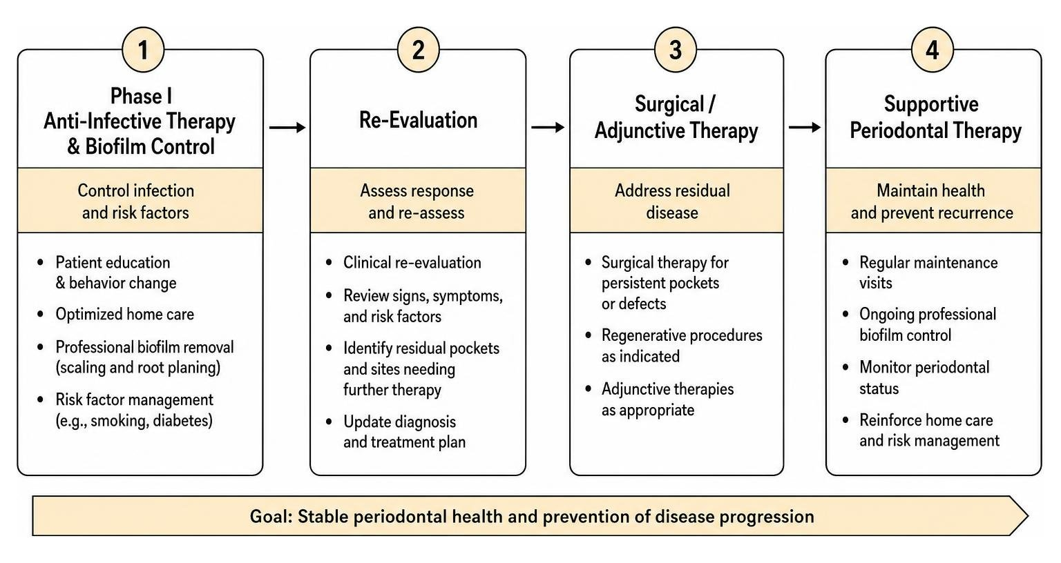
Managing periodontitis requires an organized care pathway. Modern evidence-based guidelines, such as the Treatment of stage I–III periodontitis—The EFP S3 level clinical practice guideline , establish a clear four-step clinical pathway tailored to disease severity.
Phase I Anti-Infective Therapy and Biofilm Control
Step 1 focuses entirely on behavior modification, daily home care habits, and systemic risk factor control. Without controlling external risk factors, professional treatments cannot yield lasting attachment stability.
Key components of Phase I anti-infective therapy include:
- Tailored Oral Hygiene Instruction: Training patients on proper brushing and interdental plaque removal.
- Professional Mechanical Plaque Removal (PMPR): Clearing away supragingival deposits and retentive plaque traps.
- Smoking Cessation Counseling: Tobacco use alters blood flow to the gums, suppresses immune response, and reduces healing outcomes after root planing.
- Glycemic Management: Diabetes and periodontal destruction share a bidirectional relationship. Elevated blood sugar exacerbates gum inflammation, while chronic gum infection makes blood glucose harder to regulate. Learn more about managing these interconnections in our article on the periodontal disease diabetes connection.
The Central Role of Re-Evaluation in the Periodontal Therapy Protocol
The re-evaluation phase is the critical turning point in any treatment plan. As highlighted in clinical literature such as The Trimeric Model: A New Model of Periodontal Treatment … , re-evaluation does not sit at the linear end of care; it sits at the center of all ongoing decisions.
We schedule this thorough re-evaluation 3 to 6 weeks after completing nonsurgical debridement. This window gives soft tissues enough time to form a long junctional epithelium attachment and allows soft tissue edema to subside. During this visit, we repeat 6-point probing, re-evaluate BOP, assess home care compliance, and determine whether the patient can transition to routine supportive care or requires surgical intervention.
Nonsurgical Interventions: Scaling, Root Planing, and Adjunctive Therapies
Nonsurgical periodontal therapy—also known as cause-related or etiotropic therapy—serves as the primary line of defense against active disease. Its primary aim is to decontaminate root surfaces and restore a bio-compatible microenvironment.
Scaling and Root Planing as First-Line Care
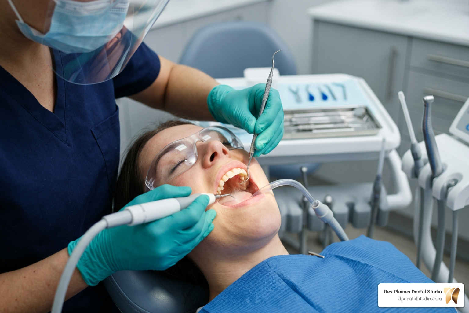
According to American Dental Association (ADA) clinical practice guidelines, scaling and root planing (SRP) is the gold standard initial therapy for chronic periodontitis because its clinical benefits significantly outweigh potential risks.
While a routine hygiene cleaning removes supragingival plaque and stains above the gumline, a therapeutic deep teeth cleaning penetrates beneath the tissue margins. Using precise hand curettes and ultrasonic scalers, we remove subgingival calculus, disrupt bacterial biofilm, and smooth rough root cementum harboring endotoxins.
Evidence-Based Use of Local and Systemic Adjuncts
While physical root debridement remains the cornerstone of care, specific chemotherapeutic or physical adjuncts may enhance outcomes in selected cases:
- Subantimicrobial-Dose Doxycycline (Periostat): Taken systemically at low doses (20 mg twice daily), this medication does not kill bacteria directly. Instead, it acts as a host modulation agent that inhibits tissue-destructive matrix metalloproteinase (collagenase) enzymes.
- Local Sustained-Release Antimicrobials: Microscopic devices placed directly inside non-responding deep pockets (>5 mm). Examples include minocycline microspheres (Arestin) delivering 1 mg of antibiotic, doxycycline hyclate gel (Atridox), or chlorhexidine gelatin chips (PerioChip).
- Systemic Antibiotics: Combination drug therapies (such as Amoxicillin paired with Metronidazole) are reserved for aggressive periodontitis or severe generalized Stage III cases in younger patients. Routine systemic antibiotic use alongside standard SRP is strongly discouraged due to antibiotic resistance risks.
- Photodynamic Diode Laser Therapy: Diode lasers may offer weak adjunctive benefits when paired with mechanical scaling, though expert consensus advises against relying on lasers as a stand-alone replacement for traditional SRP.
Indications for Periodontal Surgery and Maintenance Recall
When nonsurgical interventions fail to resolve deep pockets or structural defects, advanced surgical steps and strict recall schedules protect your long-term oral health.
When Surgical Interventions Become Necessary
The primary goal of surgical therapy (Step 3) is to establish an environment that the patient and hygienist can keep clean. Surgery is generally considered when re-evaluation reveals residual probing depths ≥6 mm or persistent deep intrabony defects.
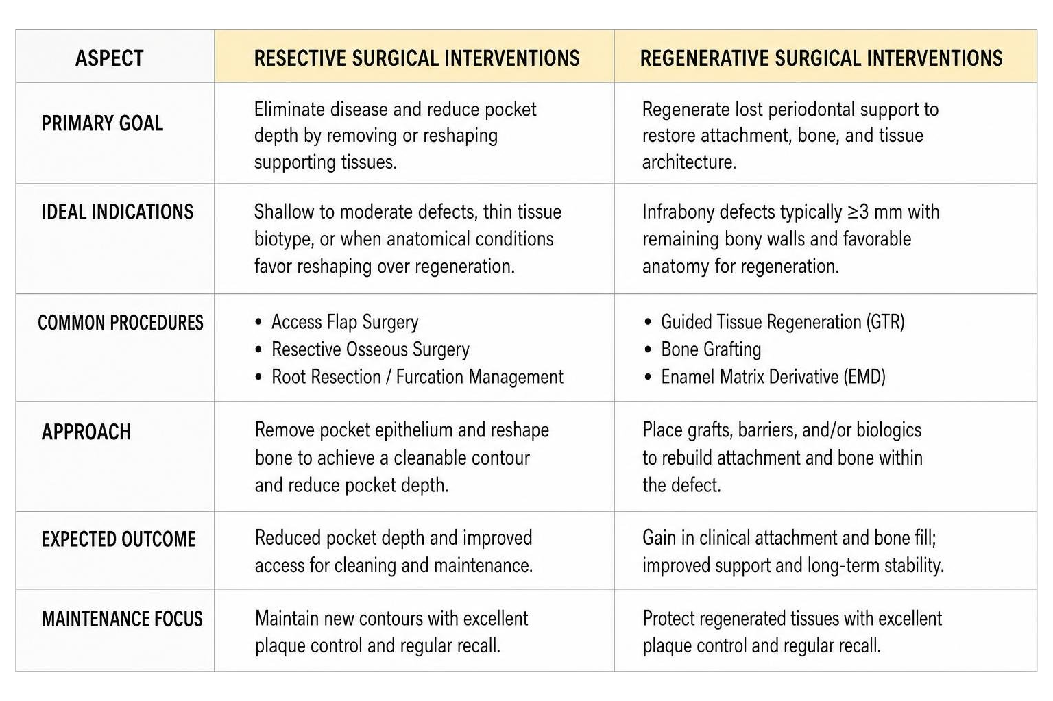
Depending on the underlying bone contour and defect type, surgical solutions include:
- Access Flap Surgery: Gently separating soft tissue to allow direct visualization and complete instrumentation of deep root surfaces.
- Resective Osseous Surgery: Reshaping uneven, cratered bone borders around teeth to eliminate deep pockets and restore smooth, natural contours.
- Regenerative Osseous Surgery: Utilizing bone grafts, barrier membranes, and tissue-stimulating proteins to rebuild lost support inside intrabony defects (typically 3 mm or deeper).
- Root Resection / Furcation Management: Treating severe class II or III furcations to clear unreachable bacterial harbors without immediately resorting to tooth extraction.
Explore our comprehensive options for surgical and advanced nonsurgical gum disease treatment.
Customizing Maintenance Schedules in a Periodontal Therapy Protocol
Once active infection is controlled, patients transition into Step 4: Supportive Periodontal Care (SPC). Skipping maintenance is one of the most common reasons periodontitis recurs. Research shows that 68% of patients with advanced disease attend only 0 to 2 maintenance visits per year in general practices—far below clinical recommendations.
Microbiological studies demonstrate that subgingival periodontal pathogens repopulate to baseline pre-treatment levels within 9 to 11 weeks following pocket instrumentation. Therefore, a 3-month recall interval (4 visits per year) is the evidence-based standard of care for patients with historical attachment loss.
During maintenance visits, we check probing depths, clean subgingival sites, review plaque control, and recommend effective home care tools. For example, tailored interdental brushes are far more effective at removing biofilm between root surfaces than standard dental floss. For more tips on keeping your mouth healthy between visits, check out our guide on providing relief from periodontal disease.
Restorative Integration, Patient Communication, and Coding Practices
Achieving long-term stability requires blending periodontal treatment with restorative care, administrative clarity, and open communication.
Interdisciplinary Care and Patient Compliance Strategies
Periodontal health is the essential prerequisite for all elective cosmetic, restorative, or orthodontic procedures. Moving teeth through inflamed bone during active infection causes rapid attachment loss. Once periodontitis is controlled, orthodontic alignment can proceed safely. Similarly, overhanging fill edges or rough crown margins harbor plaque and must be fixed during initial therapy.
Clear communication helps bridge the gap between initial diagnosis and lifelong compliance:
| Procedure Category | Dental Prophylaxis (Preventive) | Periodontal Maintenance (Therapeutic) |
|---|---|---|
| Primary Goal | Prevent onset of gum disease in a healthy mouth | Control ongoing chronic infection and prevent disease progression |
| Target Audience | Patients with healthy tissue or intact periodontium | Patients previously treated for periodontitis (SRP or surgery) |
| Subgingival Debridement | Minimal (focuses on supragingival plaque/stains) | Thorough subgingival biofilm disruption and site debridement |
| Typical Frequency | Every 6 months | Every 3 to 4 months |
| ADA Insurance Code | D1110 | D4910 |
Helping patients understand that periodontal maintenance is a lifelong therapeutic requirement—rather than an optional cleaning—improves long-term success.
Accurate Dental Coding and Documentation Practices
Accurate insurance documentation ensures treatment plans are clear and transparent:
- D0180 (Comprehensive Periodontal Evaluation): Used for full evaluations of patients showing signs of periodontal disease or risk factors.
- D4341 (Periodontal Scaling & Root Planing – 4+ teeth per quadrant): Therapeutic debridement applied per quadrant when four or more teeth exhibit pocketing and attachment loss.
- D4342 (Periodontal Scaling & Root Planing – 1 to 3 teeth per quadrant): Used for localized non-surgical therapy involving one to three teeth in a quadrant.
- D4910 (Periodontal Maintenance): Ongoing therapeutic care following active nonsurgical or surgical treatment. This code should not be alternated with D1110 (Adult Prophylaxis), as prophylaxis applies only to healthy tissues.
- D4381 (Localized Delivery of Antimicrobial Agents): Coding for sustained-release devices like Arestin inserted into stubborn pockets.
At Des Plaines Dental Studio, our staff assists patients with insurance navigation, helping maximize direct benefits while establishing care plans built around health requirements rather than annual cap restrictions.
Frequently Asked Questions About Periodontal Therapy Protocols
What is the primary difference between dental prophylaxis and periodontal maintenance?
A dental prophylaxis (ADA code D1110) is a preventive procedure designed for patients with healthy gum tissues and no history of bone loss. Its goal is to clean plaque and stains off tooth surfaces above the gumline. Periodontal maintenance (ADA code D4910) is a continuous therapeutic treatment for patients who have been treated for active periodontitis. It involves deep subgingival cleaning and pocket monitoring to prevent infection from returning. Catching issues early helps prevent advanced damage—read more in our guide on early periodontitis.
Why is a 3-month maintenance interval recommended after active therapy?
Clinical studies show that harmful periodontal bacteria start repopulating deep pockets within 9 to 11 weeks after mechanical root planing. A 3-month maintenance schedule disrupts these bacterial colonies before they can cause renewed bone destruction or attachment loss.
When are systemic antibiotics recommended during periodontal therapy?
Systemic antibiotics are not recommended for routine use during standard scaling and root planing due to risks of adverse reactions and antibiotic resistance. They are typically reserved for severe, rapidly progressing cases (such as Stage III/IV periodontitis in younger individuals) or patients with compromised immune systems. When used, systemic antibiotics must be combined directly with mechanical scaling rather than taken alone.
Conclusion

Following a standardized periodontal therapy protocol ensures that gum disease is accurately diagnosed, thoroughly treated, and carefully monitored over time. By combining initial nonsurgical therapy like scaling and root planing with personalized maintenance care, the vast majority of patients can stop disease progression and retain their natural teeth for a lifetime.
At Des Plaines Dental Studio, we deliver gentle, evidence-based care tailored to your unique smile. If you have questions about your gum health or want to schedule an evaluation, reach out to our team today to explore our dedicated options for periodontitis treatment.